Female | 32
Is my liver size and echotexture normal?
LIVER: Is normal in size (15.5 cms) and echotexture. No focal lesions are seen. No dilatation of intra-hepatic biliary radicles present. Portal vein is normal. Common bile duct is normal. GALL BLADDER: Is distended. Normal in wall thickness. No calculus or mass. PANCREAS: Visualized head and body appears normal. Rest obscured by bowel gas SPLEEN: Is normal in size (9.9 cms) and echotexture. RIGHT KIDNEY: Measures 9.2 * 3.7 cms. Normal in size and echotexture. Cortico medullary differentiation is well maintained. No calculus, hydronephrosis or mass. LEFT KIDNEY: Measures 9.9 * 3.6 cms. Normal in size and echotexture. Cortico medullary differentiation is well maintained. No calculus, hydronephrosis or mass. URINARY BLADDER: Is distended. Normal wall thickness. Few echogenic particles noted in the lumen. No obvious calculus or mass. No vesical diverticulum present. UTERUS Measures 8.3 * 4.3 * 5.8 cms. Normal in size. Small hypoechoic lesion of size 8.5 * 5.5 mm noted involving the posterior myometrium - possibly fibroid. Endometrial thickness measures 5.6 mm Right ovary measures - 52.7 * 19.6 * 42.2mm volume- 22.8 cc Left ovary measures - 45.5 * 23.2 * 44.4 mm, volume - 24.5 cc Both ovaries are slightly bulky in size and shows mild increase in stromal echoes with multiple small follicles of size 3-5mm. No dominant follicle noted in either side. No adnexal mass lesion seen. No free fluid in POD. Both iliac fossae appear normal and there is no obvious evidence of bowel mass or bowel wall thickening present. IMPRESSION: Few echogenic particles in the urinary bladder lumen. Suggested urine routine correlation Small uterine fibroid. Polycystic appearance in both ovaries. Suggested follow up & clinical correlation

Gynecologist/Obstetrician
Answered on 7th May '24
The results show you may have a small growth called a fibroid in your uterus. This is not cancer. But it can cause heavy periods or pain in your lower belly. The results also show some cysts on both ovaries. This is called polycystic ovaries. With this condition, your periods may be irregular or you may have trouble getting pregnant. To understand better, you should take a urine test and visit a gynecologist. With proper care from your doctor, you can manage these issues well.
Related Blogs
What is Intrauterine Insemination (IUI)?
Intrauterine insemination (IUI) is also known as artificial insemination. Get all the details about IUI treatment with the complete process, uses and risks.

10 Best Hospitals in Istanbul - Updated 2023
Looking for the best hospital in Istanbul? Here is a compact list for you of the 10 Best Hospitals in Istanbul.
Labiaplasty Turkey (Compare costs, clinics & surgeons 2023)
Experience labiaplasty in Turkey. Explore skilled surgeons and state-of-the-art facilities for safe, confidential, and personalized procedures tailored to your needs and desired outcomes.

Dr. Hrishikesh Dattatraya Pai- Fertility Specialist
Dr. Hrishikesh Pai is a highly experienced gynecologist and obstetrician pioneering many assisted reproductive technologies in India to help couples fight infertility and achieve pregnancy.
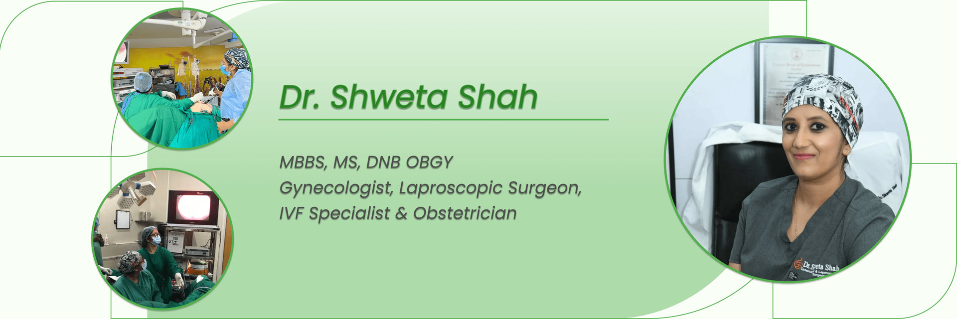
Dr. Shweta Shah- Gynaecologist, IVF Specialist
Dr. Shweta Shah is well-renowned Gynaec, Infertility Specialist, and Laparoscopic Surgeon who has medical working experience of 10+ years. Her area of expertise is a high-risk pregnancy and invasive surgery related to women's health problems.