Asked for Male | 53 Years
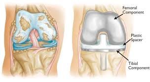
I am 53 yrs old recently I had surgery from fortis hospital Post operative appearance with orthopedic device, plates and screws seen in situ. There is evidence of a persistent fracture line in the distal femoral condyle with extensions involving patellofemoral articular surface, intercondylar notch, medial and lateral condyle reaching tibiofemoral articular surface. Proximal visualized shaft of left femur shows diffuse cortical thickening, coarse trabeculation and patchy intramedullary sclerosis. Proximal end of fracture does not show any clearly demonstrable callus formation or periosteal reaction suggesting hypo/oligotrophic fracture healing. Multiple well defined small bony hyperdensities are seen within the fracture line. Extensive surrounding soft tissue stranding and fluid density seen within the intercondylar notch area. Osteoarthritic changes seen involving the knee joint with tibial spiking, marginal osteophytes, significantly reduced medial tibiofemoral joint space.
Answered by Dr. Rajat Jangir
We would be more than happy to help you but you have just told your reports but did not mention What's your problem? So kindly get in touch with an orthopedic surgeon near you.

Joint Replacement Surgeon
Questions & Answers on "Orthopedic" (1356)
Related Blogs

Painless knee-replacement in India
Here is all the information you need to know about painless knee replacement (Minimally Invasive Surgery) in India.

Overweight and Obesity: Understanding Health Impacts
Confronting overweight and obesity. Explore causes, risks, and effective strategies for achieving a healthier lifestyle. Take control today!

Hip Replacement Hospitals in India: A Comprehensive Guide
Hip pain slowing you down? Transform your mobility with India's top-rated Hip Replacement experts. Experience minimally invasive surgery, affordable costs, exceptional outcomes, cutting-edge technology, compassionate care, & proven results await!

10 Best Knee Replacement Hospitals in India
Unlock mobility and reclaim your life with leading knee replacement hospitals in India. Experience expert care, state-of-the-art facilities, and affordable solutions for your needs.

When physiotherapy is not the only option left...
Here are all the things you should know before getting a knee replacement in India
Cost Of Related Treatments In Country
Acl Reconstruction Cost in India
Bone Densitometry Cost in India
Rheumatoid Arthritis Treatment Cost in India
Spine Surgery Cost in India
Limb Lengthening Cost in India
Spinal Muscular Atrophy Cost in India
Spinal Fusion Cost in India
Arthroscopy Cost in India
Slip Disc Cost in India
Hip Replacement Cost in India
Top Different Category Hospitals In Country
Heart Hospitals in India
Prostate Cancer Treatment Hospitals in India
Kidney Transplant Hospitals in India
Cosmetic And Plastic Surgery Hospitals in India
Dermatology Hospitals in India
Endocrinology Hospitals in India
Gastroenterology Hospitals in India
Gynaecology Hospitals in India
Hematology Hospitals in India
Hepatology Hospitals in India
Top Doctors In Country By Specialty
Top Orthopedic Hospitals in Other Cities
Orthopedic Hospitals in Chandigarh
Orthopedic Hospitals in Delhi
Orthopedic Hospitals in Ahmedabad
Orthopedic Hospitals in Mysuru
Orthopedic Hospitals in Bhopal
Orthopedic Hospitals in Mumbai
Orthopedic Hospitals in Pune
Orthopedic Hospitals in Jaipur
Orthopedic Hospitals in Chennai
Orthopedic Hospitals in Hyderabad
Orthopedic Hospitals in Ghaziabad
Orthopedic Hospitals in Kanpur
Orthopedic Hospitals in Lucknow
Orthopedic Hospitals in Kolkata
- Home >
- Questions >
- I am 53 yrs old recently I had surgery from fortis hospital ...