As we delve into the stage 4 esophageal cancer survival rate by age, it’s crucial to study the record in the most recent global data available.
In 2024, esophageal cancer remains the eighth most common cancer worldwide, with over 600,000 new cases reported in 2020. The survival rates for this formidable disease vary significantly with age. For those diagnosed with stage 4 esophageal cancer, the overall five-year survival rate hovers around 20%. But this number declines as age increases.
As we explore the intricacies of stage 4 esophageal cancer survival rates by age, we aim to shed light on the factors behind these numbers, the advancements in medical science striving to improve them, and the collective effort needed to change the course of this disease.
What is Stage 4 Esophageal Cancer?
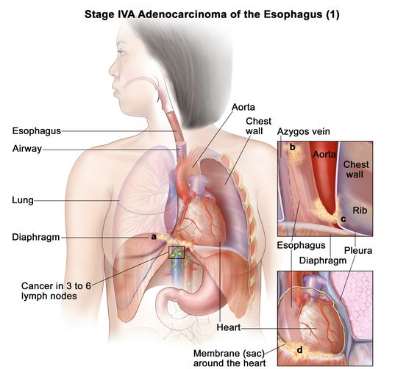
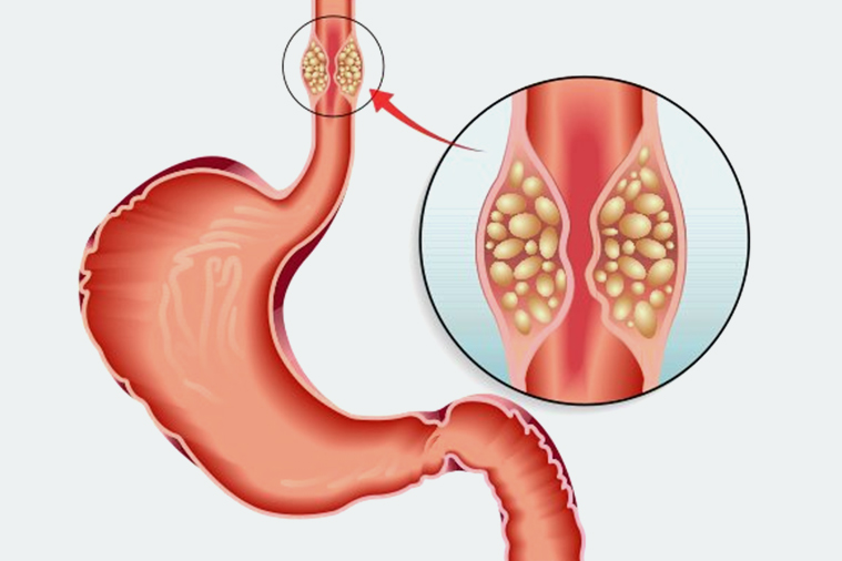
Stage 4 esophageal cancer refers to cancer that has spread beyond the esophagus to other parts of the body. This stage is considered advanced and typically involves regions such as nearby lymph nodes or distant organs, including the liver or lungs. The prognosis for this stage is generally poor, but various factors can affect individual outcomes.
If you or a loved one has been diagnosed with esophageal cancer, schedule a consultation with the experts to discuss your specific case and the best treatment options available for you.
Survival Rates by Age Group for Stage 4 Esophageal Cancer
As of 2023, the overall five-year survival rate for esophageal cancer is around 20%, but it varies significantly by age group:
- The five-year survival rate for individuals younger than 55 is approximately 27%.
- The survival rate for those aged 55 to 64 drops to about 17%.
- The survival rate for those 65 or older decreases to around 11%.
- Patients with stage III/IV esophageal cancer under the age of 45 had 1-year, 3-year, and 5-year survival rates of 88.6%, 31.8%, and 24.2%, respectively.
- In comparison, the older matched cohort (above 45) had 1-year, 3-year, and 5-year survival rates of 58.3%, 20.6%, and 8.8%
It’s crucial to note that these statistics represent average, and individual outcomes vary widely based on various factors, including the patient’s overall health, response to treatment, and medical advancements.
What does a 5-10% survival rate indicate?
A 5-10% survival rate suggests that only 5 to 10 out of every 100 patients are expected to survive for a specified period (usually five years post-diagnosis). This low percentage highlights the aggressive nature of stage 4 esophageal cancer and its poor prognosis.
Factors Affecting Survival Rates
- Age: Younger patients often have better survival prospects.
- Overall Health: Stronger overall health enables tolerating aggressive treatments.
- Tumor Characteristics: The location and size of the tumor influence treatment outcomes.
- Cancer Type: Different cell types respond differently to treatments.
- Cancer Spread: Extent of metastasis critically impacts survival rates.
- Treatment Quality: Access to advanced treatments can improve survival.
- Genetics: Certain genetic factors affect treatment effectiveness.
- Lifestyle: Smoking, alcohol, and nutrition can alter disease progression.
- Treatment Response: Initial success with treatment typically extends survival.
What is the longest survival rate for esophageal cancer?
The overall five-year survival rate for esophageal cancer is about 20%, but survival rates can range from 5% to 47%. When esophageal cancer is found early and when it is small, the five-year survival rate is higher.
What if cancer has spread to the lungs?
If esophageal cancer has spread to the lungs, the condition is classified as metastatic and the treatment becomes more complex and palliative. The focus shifts to managing symptoms and improving quality of life rather than curative intent.
For more detailed information about esophageal cancer and treatments, Book an appointment with our oncologists to understand your diagnosis and the path forward.
Treatment Options for Stage 4 Esophageal Cancer
- Chemotherapy: Often the first line of treatment to shrink tumors and manage symptoms.
- Radiation Therapy: Used to relieve pain and other symptoms, sometimes in combination with chemotherapy.
- Targeted Therapy: Focuses on specific genetic markers of cancer cells to block their growth.
- Immunotherapy: Boosts the immune system's ability to fight cancer, especially for tumors with certain genetic characteristics.
- Palliative Surgery: Aims to relieve symptoms but not to cure, such as stenting to keep the esophagus open.
- Supportive Care: Includes nutrition support, pain management, and other therapies to improve quality of life.
Conclusion
While the prognosis for stage 4 esophageal cancer can be daunting, understanding the survival rates by age and the available treatments can help patients and caregivers make informed decisions. It is vital to discuss all possible treatment options with a healthcare provider.
FAQs:
Does esophageal cancer spread quickly?
Yes, esophageal cancer can spread quickly compared to other cancers, particularly if it starts in the cells lining the esophagus.
Can stage 4 esophageal cancer be cured?
While stage 4 esophageal cancer is rarely curable, treatments can help alleviate symptoms.
Is esophageal cancer painful?
Yes, esophageal cancer is often painful, especially as it progresses. Pain may arise from the tumor itself or as a side effect of treatments like chemotherapy or radiation.
References:


